How Ultrasound Guided Injections Improve Filler Outcomes
8 May 2025
In this post:
- Ultrasound guided injections offer real-time visualisation of anatomy, enabling safer, more precise filler placement and reducing the reliance on guesswork.
- Blind injecting increases the risk of complications such as vascular occlusion, asymmetry, or poor outcomes.
- Ultrasound enhances clinical practice by allowing injectors to verify filler placement, manage complications, and build trust with patients.
- Training in facial ultrasound is essential to use the tool effectively, and the Smileworks Aesthetic Training HUB offers comprehensive courses to help injectors integrate ultrasound into their daily aesthetic practice.
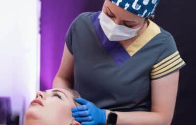
When a patient chooses to put their health and wellbeing in your hands, precision and safety are not a compromise. They are a clinical necessity.
As the demand for non-surgical aesthetic treatments grows, so does the pressure on practitioners to deliver safe, effective, and tailored results. Currently, injecting into the skin ‘blind’ is the industry norm. This relies heavily on anatomical averages and practitioner experience. This makes it risky for new and less experienced injectors.
Every face is different, and every layer beneath the skin needs to be considered when putting a needle or cannula into the skin. With blind injections, there is no way to do this properly. It’s mostly down to guesswork, even from the most experienced of injectors. This is where ultrasound guided injections become useful.
In this blog post, we’ll explore how facial ultrasound enhances safety, ensures proper filler placement, and why it’s an absolute must for any injector, regardless of experience.
What Are Ultrasound Guided Injections?
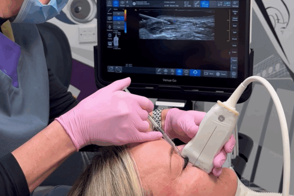
Ultrasound guided injections in aesthetics utilise high-frequency ultrasound imaging, typically in a linear transducer in the 15-22 MHz range. This allows you to visualise soft tissues, blood vessels, fascia, and filler material in real time.
You’ll be able to:
- Map vascular and structural anatomy before injecting
- Visualise needle or cannula movement during filler placement
- Confirm exact filler location immediately post-injection
- Diagnose and manage complications with target intervention
This level of insight replaces educated guesswork with anatomical evidence. It is without a doubt the safest way to inject. You’ll no longer have to estimate the location of key structures like the facial artery, angular artery, deep fat compartments, and fascial planes. The ultrasound imaging will show you the precise anatomy in real-time.
Ultrasound is also a standout aesthetic tool for its ability to distinguish between tissue layers, vascular structures, and filler deposits. This opens the door to safer, more thorough aesthetic treatments, even in complex cases.
The Problem With ‘Blind’ Injecting
You’ll find that most clinics still rely on blind injection techniques based on textbook anatomy and surface landmarks. While experienced injectors often develop excellent tactile awareness and intuitive spatial judgment over time, there are still several critical limitations with blind injecting.
Most reported side effects are minor and temporary. These commonly include:
Anatomical Variability
Facial anatomy in aesthetics varies not just between individuals, but also between sides of the same face. Arteries can follow unexpected paths. Fat compartments may be asymmetric. Patients with prior filler, trauma, surgery, or scarring may present unpredictable tissue planes. Blind injecting ignores this variability, increasing the chances of suboptimal outcomes or complications.
Increased Risk in High-Stakes Areas
Zones such as the glabella, nose, tear trough, and temple contain dense vascular networks and critical end arteries. Injecting in these areas without imaging can increase the risk of:
- Intravascular injection → vascular occlusion → ischemia, necrosis, and/or filler blindness
- Compression of key arteries by poorly placed filler
- Unintended retrograde embolisation leading to vision loss
These risks, although rare, are potentially devastating. A single case of botched tear trough filler can be the end of your aesthetics career.
Difficulty Assessing Old Filler
Ultrasound is imperative for conducting facial assessments, especially in patients with previous treatments often present migrated, nodular, or deep seated filler. Sometimes this can be years old. Without imaging, there’s no way to distinguish between tissue and filler material, complicating correction or retreatment. Ultrasound provides a non-invasive, real-time method to assess and plan safe revision procedures.
Clinical Benefits of Ultrasound Guided Injections
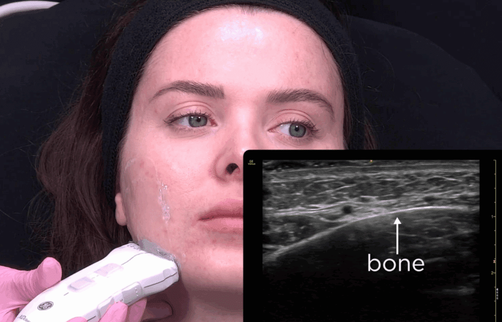
1. Anatomical Precision
Ultrasound allows direct visualisation of skin depth, tissue layers, and vascular structures. This real-time mapping means:
- Cannulas or needles are guided around arteries, not through them
- Fillers are placed at the intended plan (e.g., supraperiosteal vs. subcutaneous)
- Fat compartments and retaining ligaments are targeted accurately for structural support
This is particularly valuable in complex areas like the midface or with temple filler, where mere millimetres make a massive difference. A more anatomically tagged approach translates to a better result for your patient.
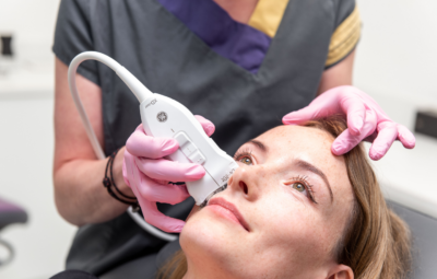
For a preview into our Facial Ultrasound course, see this preview where HUB lead instructor and founder Dr MJ Rowland-Warmann guides you through how to identify key anatomical structures using ultrasound.
2. Filler Placement Verification
With ultrasound, injectors can observe the echo texture of the filler and confirm its location immediately. This ensures:
- Even, symmetrical placement
- Avoidance of overfilling or superficial boluses
- Elimination of unintended intramuscular or intravascular filler
This immediate verification can prevent aesthetic irregularities such as lumps, asymmetries, or the Tyndall effect. It also reassures both you and your patient that the product has gone exactly where it was supposed to.
3. Real-Time Complication Management
In cases of suspected vascular occlusion, ultrasound can:
- Detect filler that has blocked a vessel
- Guide hyaluronidase precisely to affected areas
- Monitor reperfusion and tissue response
Ultrasound is equally useful for treating delayed-onset nodules, granulomas, or Tyndall effect. You’ll be able to identify the cause, plan targeted treatment, and verify resolution. This leads to quicker intervention and better clinical outcomes.
4. Higher Patient Trust and Professional Differentiation
In an industry that’s so unregulated, more and more patients today expect transparency, accountability, and reassurance. The ability to show them their anatomy on screen, explain why certain areas are high-risk, and demonstrate post-treatment filler placement increases trust and sets you apart clinically. Facial ultrasound will set you apart as having a commitment to safety and excellence.
It also aligns with growing patient interest in technology-driven care and precision aesthetics. Too often have they seen cases of lip filler gone wrong; they want safe, evidence-backed treatments. Patients will look at facial ultrasound not only as a safety tool but also a marker that your practice is modern and high-end.
Common Clinical Use Cases
Here’s where ultrasound guided injections are proving most impactful:
- Tear Troughs: Identify the infraorbital artery and verify sub-orbicularis oculi placement to reduce risk of puffiness or vascular occlusions
- Nasal Filler: Avoid dorsal nasal arteries and confirm deep periosteal filler placement
- Temple Hollowing: Navigate the superficial temporal artery and temporalis fascia with clarity
- Lip Revision: Detect and dissolve lip filler from past treatments
- Emergency Management: Use Doppler ultrasound to assess vessel patency post-injections if complications arise
Even in routine, easy-to-inject areas like the cheeks, ultrasound enhances confidence and individualisation. It allows you to tailor injection depth, volume, and technique to your patient’s unique anatomy. Every patient will receive a bespoke treatment tailored to their individual anatomy rather than a one-size-fits-all injecting approach.
Important Considerations
Ultrasound is powerful, but it’s not simply a plug-and-play tool. There are many factors that will contribute to your personal success with the tool.
- Technical Training: Learning probe orientation, image interpretation, and needle tracking takes time. A structured ultrasound course and mentoring sessions are essential.
- Anatomical Fluency: Sonographic anatomy looks different from textbook illustrations. You need to re-learn landmarks in grayscale.
- Equipment Investment: Ultrasound devices from brands like Clarius or GE offer high-resolution imaging but come at a cost. However, the ROI in clinical quality and complication avoidance is high.
- Workflow Evolution: Initially, treatments may take longer as you build scanning proficiency. However, with experience, ultrasound becomes an intuitive part of the assessment and injection process
The learning curve is real, but so is the reward. With the right support and systems in place, facial ultrasound can integrate seamlessly into your practice.
Why More Practitioners Are Adopting It
In the UK, the aesthetic industry remains notoriously unregulated. Year after year, complications only become more commonplace. This only increases the need for improved clinical governance, documentation, and patient safety protocols. Luckily, ultrasound aligns perfectly with these priorities:
- Improves documentation with scan images before and after treatment
- Enhances medico-legal defensibility
- Boosts patient safety metrics
- Supports CPD and ethical development
Practices that adopt ultrasound are seen as progressive, evidence-based, and patient-first. In the competitive world of aesthetic medicine, this can significantly improve your positioning with current and future patients.
We’re also seeing patient awareness of the risks and complications of injectable treatments grow. Clinics that can demonstrate proactive risk management and transparency are more likely to retain loyalty and generate referrals.

How to Get Started with Ultrasound Guided Injections
It’s time to properly introduce ourselves. We are the Smileworks Aesthetic Training HUB – an esteemed aesthetic training centre based in the UK. We pride ourselves on our wide range of online and hands-on courses tailored for every skill level.
At the HUB, we offer the world’s most comprehensive training pathways in aesthetic ultrasound. Facial ultrasound is not just a tool for rare complication cases or advanced injectors. It’s the foundation for a safer, smarter, and more ethical practice.
For top-tier ultrasound learning, look no further than our Foundation Facial Ultrasound Course and our Advanced Facial Ultrasound Course, two musts for all injectors. Ultrasound is the single most important tool for ensuring safe and effective treatments every time.
For those looking for more in-depth training, we recommend booking a one-to-one mentoring session with Dr MJ. These sessions at 100% customisable, and you’ll receive personalised instruction based on your skill level and goals.
Join us at the HUB. Let’s shape the future of aesthetic medicine together and ensure safe, effective, and transformative results for every patient.
Want to try out our courses before committing? Take advantage of our free trial for a taste of what learning at the HUB is like.
Related blog posts:
Remarkable Aesthetics Courses
Advanced Facial Ultrasound
Explore more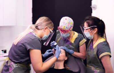
One-to-One Mentoring
Explore more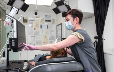
Facial Ultrasound
Explore more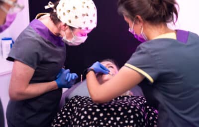
Lip Filler
Explore more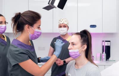
Botox
Explore more